Abstract
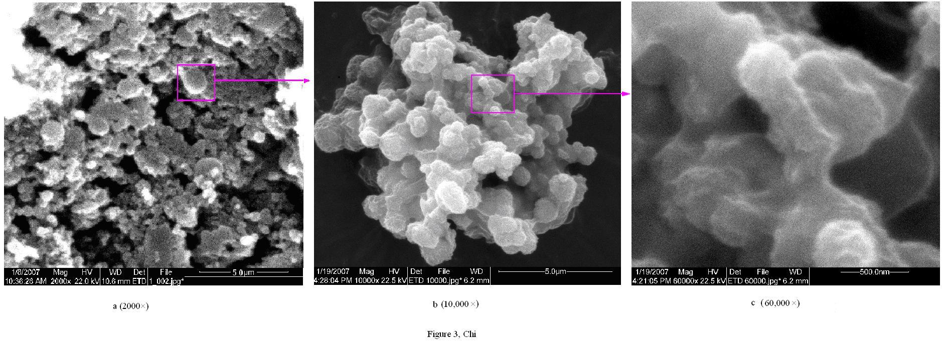
The crude polysaccharide was obtained from Gynostemma pentaphyllum Makino by water extraction followed by ethanol precipitation. The polysaccharide was successively purified by chromatography on DEAE-52 and SephadexG-150 column, and three polysaccharide fractions were obtained and termed GPP1-a, GPP2-b and GPP3-a, respectively. The administration with GPP1-a prolonged markedly exhaustive exercise time of the mice. Structural features of GPP1-a were investigated by a combination of instrumental and chemical analysis, such as atomic force microscope (AFM), scanning electron microscope (SEM), partial acid hydrolysis, periodate oxidation, Smith degradation, methylation analysis, gas chromatography-mass spectrometry (GC-MS) analysis and NMR spectroscopy. The results indicated that GPP1-a has a backbone of (1→4)-linked α-D-Glucose residues, which occasionally branches at O-6. The branches were mainly composed of (1→6)-linked α-D-Glucose, (1→3)-linked β-D-Galactose and (1→6)-linked α-D-Galactose residues, and terminated with β-D-Galactose residues and β-L- Arabinose residues.
Keywords: Gynostemma pentaphyllum Makino; Polysaccharide; Anti-fatigue; Morphology; Structure
1. Introduction
It is well accepted that carbohydrates can serve as structural components and energy source in the cell. Their highly complex structure allows very specific interactions, so that these biomolecules are involved in a variety of molecular recognition processes in intercellular communication and signal transductions such as cell adhesion, differentiation, development, regulation, etc. (Varki, 1993; Wang, et al., 2007).
As we all have known that chemical structure of polysaccharides is the base of its bioactivity, for example, polysaccharides from the different strains of Poria cocos mycelia showed different in vivo and in vitro anti-tumor activities, depending on their monosaccharide composition, molecular mass, and chain conformation (Jin, et al., 2003). A more recent study has shown that the different monomers ingredients from the same crude polysaccharide have different biological activities (Tiffany, et al., 2007). Therefore, it is valuable that the isolation and characterization of the monomers ingredient with strong biological activity from crude polysaccharide is beneficial for their usage in the prevention and treatment of related-diseases, such as anti-tumor.
It has been found that the structure of polysaccharide is intimately related to its functions. This notion was further supported by the study indicating that the majority of polysaccharide from mushrooms with anti-tumor activity has β (1→3)-D-glucan structure in the main chain (Akira, et al., 1981). Glucan with three helix structure and molecular weight of more than 90,000 enhances immune functions (Ohno, et al., 2001), indicating that molecular morphology of polysaccharide was closely associated with its functions. Therefore, the structural identification of polysaccharides is necessary for better understanding of the relationship between structure and activity.
Gynostemma pentaphyllum Makino is a kind of perennial herbs climbing shrub of Gynostemma pentaphyllum genus of gourd family, and it mainly grows in oriental countries (Hu, et al., 1996). Numerous studies on Gynostemma pentaphyllum Makino have illustrated a variety of biological activities with minimal pharmacological toxicity (Attawish, et al., 2004), including anti-gastric ulcer (Rujjanawate, et al., 2004), anti-tumour (Li, et al., 1993), enhancing immune function (Zhou, et al., 2006) and cardiovascular protection (Circosta, et al., 2005). To data, its biological activities were mainly attributed to gypenosides. However, recent studies have suggested that the polysaccharide components of Gynostemma pentaphyllum Makino also exhibits many activities including anti-aging (Luo, & Wang, 2005), anti-oxidant activities (Wang, & Luo, 2007) and improving immune competence (Qian, et al., 1999). Moreover, the crude polysaccharide from Gynostemma pentaphyllum Makino enhanced exercise capability in mice (Fu, 2000). These results clearly demonstrated that polysaccharide is also an active component of Gynostemma pentaphyllum Makino. However, whether polysaccharide is consisted of several distinct fractions remain elusive. More important, the structural and morphological information of the fraction of polysaccharide with the strongest biological activities molecular was lacking.
The objective of this study was to isolate the anti-exercise fatigue fractions of polysaccharide from Gynostemma pentaphyllum Makino, and to further characterize its structure and morphology.
2. Materials and methods
2.1 Materials
Gynostemma pentaphyllum Makino was purchased from Shaanxi Province (China). The columns of DEAE-52 cellulose, SephadexG-150 and Sephadex G-200 were from Pharmacia (Sweden). Standard dextrans were from Sigma. All other reagents were of the highest available quality in China.
2.2 Isolation and purification of polysaccharides
The Gynostemma pentaphyllum Makino (500 g) was extracted in 80% ethanol at 50 ºC for 2 h twice, then it was dried and extracted with distilled water at 80 ºC for 2 h twice (Solid-liquid ratio is 1:15). After each extraction, the polymers were separated from residues by filtration, and extracts were combined, concentrated and removed free protein layer by the use of method of Sevage (repeat 7 times), then concentrated and dialyzed against running water for 48 h. The above-mentioned extract was subjected to the precipitation with four-fold volumes of ethanol. The Gynostemma pentaphyllum polysaccharide (GPP) was collected by centrifugation, washed successively with ethanol, acetone and ether, and freeze-dried.
DEAE-52 cellulose column chromatography was used for the fractionation of this preparation. The sample GPP (600 mg) was dissolved in 10 mL distilled water, centrifuged, and the supernatant was injected into a column (2.5 × 60 cm) of DEAE-52 cellulose equilibrated with distilled water. After loading with sample, the column was eluted with 300 mL of distilled water at rate of 0.8 mL/min, followed by the stepwise elution using 300 mL of NaCl aqueous solution (from 0 to 1 mol/L) at rate of 0.6 mL/min, then sediment was collected with automatic fraction collector and detected by the phenol–sulfuric acid method (Dubois, et al., 1956), and each eluting peak was recorded, dialyzed against tap water and distilled water for 48 h, and then purified by gel-filtration chromatography on a column of Sephadex G-150 (2.5 × 80 cm). The sample was dissolved in the minimal volume of 0.1 mol/L sodium chloride solution and added to the column, then eluted with 0.1 mol/L solution. The main fraction is collected as described above.
2.3 Purification determination and general chemical properties of the polysaccharides
Purification of the polysaccharides was conducted using a Sephadex G-200 column chromatography and methods of optical rotation (Wang, et al., 2004). The sample were dissolved in 0.1 mol/L sodium chloride, centrifuged, then the supernatant was applied to a Sephadex G-200 column (1 × 80 cm), which was eluted with 0.1 mol/L sodium chloride at a rate 0.2 mL/min. Polysaccharides were detected by the phenol-sulfuric acid method. Elution curve was drawn by tube number as abscissa and absorbance as vertical coordinate. In addition, the sample was dissolved in distilled water, and its optical rotation was measured with a WZZ-2SS polarimeter (Xi'an Instrument Co., Xi'an, China) at 20 ºC.
The molecular weight was calculated by the calibration curve obtained by using various standard dextrans (Wang, et al., 2001). Gas chromatography (GC) was used for identification and quantification of monosaccharide composition (Wang, & Luo, 2007). The sample was carried out using the trimethylsilylation reagent. GC was then performed on a 6890 N instrument (Agilent Technologies Co., USA) equipped with capillary column (30 m × 0.32 mm × 0.25 µm). The operation was performed using the following conditions: H2: 20 mL/min; air: 200 mL/min; N2: 20 mL/min; injection temperature: 250 ºC; detector temperature: 250 ºC, the column temperature was kept at 150 ºC for 2 min, increased to 190 ºC at rate of 7 ºC/min and held for 2 min at 190 ºC, then increased to 260 ºC at rate of 7 ºC/min and held for 2 min at 260 ºC.
The infrared (IR) spectrum of the sample was determined using the KBr-disk method with a Fourier transform Infrared Spectrometer (Brucher Corp., Germany) in the range of 400 cm-1 to 4000 cm-1 (Kumar, et al., 2004). The UV spectrogram of samples was measured from 190 nm to 600 nm of wave length with a Lambd 950 instrument (Perkin-Elmer Corp., U.S.A.).


 最新动态
最新动态